Blogs

Jurassic World Evolution Becomes Jurassic Park

Sunday, November 17, 2019

DLC Brings Jurassic Park to Jurassic World Evolution

Developers Frontier Developments created an immersive park builder inspired by Jurassic World and now gamers will be able to recapture the magic of the classic Jurassic Park films in DLC releasing next month. “Return to Jurassic Park,” for Jurassic World Evolution allows gamers to create dinosaurs, locations, buildings and more inspired by the first Jurassic Park movie. The DLC will take place on the islands of Isla Nublar and Isla Sorna that the first movie was located on. “Return to Jurassic Park” releases on December 10 for $19.99 on PS4, Xbox One and PC.

Apparently, in addition to the new in-game content, the “Return to Jurassic Park” DLC also features brand new story missions featuring Drs. Alan Grant, Ellie Satler, and Ian Malcolm, who will all be voiced by the original actors from the film: Sam Neill, Laura Dern, and Jeff Goldblum, respectively. The upcoming DLC gives gamers of Jurassic World Evolution a strong new experience with lots of nods and references to the iconic film that started it all. Many gamers purchased Evolution to fulfill their dreams of living in the original park but now gamers will be able to simulate that experience in a more organic way.

We’re looking forward to making new parks in the “Return to Jurassic Park” DLC as that’s where our love of the Jurassic film franchise came from. We hope that none of our guests experience the unfortunate bathroom dilemma that Nedry did or the nail biting encounter with a bloodthirsty T-rex like the kids of Jurassic Park. Are you curious what we thought about the detailed dinosaur park simulator known as Jurassic World Evolution?

Are you excited to build your own unique Jurassic Park on the islands that kicked off the franchise or are you content building in the Jurassic World universe? Which of the Jurassic films is your favorite? Let us know in the comments below!

Source: Dualshockers / https://cogconnected.com/

Gorgeous Collection of Fossil Feathers Reveals How Dinosaurs Kept Warm in Ancient Gondwana

Sunday, November 17, 2019

(Kundrát et al., Gondwana Research, 2019)

Life for dinosaurs living at the South Pole wouldn't have been easy. Sure, it wasn't the icy hellscape it is today, but the long, dark winters still would have been frosty. Now, we have a better idea of how at least some of these animals stayed warm.

A team of scientists from Slovakia, Sweden, Australia, and the US has analysed fossils representing an array of feathers grown by dinosaurs and birds that once lived within the southern polar circle.

While hints of lush feathery dinosaurs have popped up in the fossil record here and there, most examples come from the Northern Hemisphere, representing an array of coverings that could have helped Mesozoic wildlife regulate their temperatures, hide, and occasionally even glide in relatively warm climates.

"Yet, to date, no directly attributable integumentary remains have been discovered to show that dinosaurs used feathers to survive in extreme polar habitats," says Benjamin Kear, a palaeontologist from Uppsala University in Sweden.

That's not to say we're yet to find a single feather fossil in the Southern Hemisphere. A dig site in Australia's southern state of Victoria has given up a few notable examples over the decades. They've just never been looked at closely until now.

"Fossil feathers have been known from Koonwarra since the early 1960s, and were recognised as evidence of ancient birds, but have otherwise received very little scientific attention," says Thomas Rich of the Melbourne Museum in Australia.

"Our study is thus the first to comprehensively document these remains, which include new specimens that were examined using cutting-edge technologies."

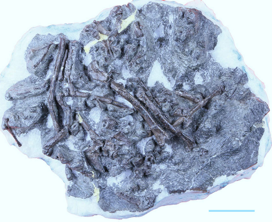
A total of ten fossil specimens were included in the study, all dated around 118 million years old, providing solid evidence of wing feathers from ancient birds, tufted dinosaur 'protofeathers', and even partly decomposed body feathers.

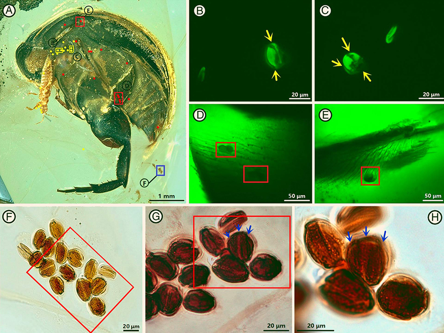
The technologies included advanced forms of microscopy and spectroscopy, allowing the team to capture an impressive level of detail from the well-preserved remains, providing information on their anatomy and – in some cases – colouration.

Some of the feathers were relatively advanced, sporting barbed 'zips' similar to modern feathers that helps them interlock for flight, and gives the animal protection against the elements.

But it was the simpler 'fluffy' feathers, like those below, that were of particular interest.

(Kundrát et al., Gondwana Research, 2019)

"Dinosaur 'proto-feathers' would have been used for insulation," says lead author Martin Kundrát, of Pavol Jozef Safarik University in Slovakia.

"The discovery of proto-feathers at Koonwarra therefore suggests that fluffy feather coats might have helped small dinosaurs keep warm in ancient polar habitats."

To understand the conditions these dinosaurs lived in, we need to rewind the clock a few hundred million years, when Earth's familiar map of continents would have looked rather different.

Today's southern landmasses - Antarctica, Australia, South America, and Africa, along with India and Arabia – were all mushed together in one giant supercontinent called Gondwana, which sat more or less directly centred on Earth's South Pole.

The world's climate was a lot warmer back then and Gondwana wasn't a winter wonderland all year round. Instead it was far more temperate, with lush ecosystems full of plants and animals.

While it wasn't exactly freezing, the poles still experienced long periods of sunlight in summer, and darkness in winter. So anything living in such extreme conditions still had to deal with an extended, chilly twilight.

Having hard evidence of potentially insulating feathers helps researchers fill in the missing pieces.

The team also found densely packed fossil melanosomes, or pigment bodies, that indicated dark colouration that might help to absorb heat, if not also help with camouflage or communication in those dimly lit months.

Ten years ago, Australian palaeontologists found clear evidence of a 110 million year old 'dino-burrow' in another Victorian dig site, suggesting at least some species could have gone underground to wait out the cold.

There's no doubt a rich variety of features and behaviours that would have helped polar animals live comfortably in such a variable environment; and it's nice to finally have some firm evidence of them.

This research was published in Gondwana Research.

Source: www.sciencealert.com/

'Friends': How Much Did Ross Geller Make as a Paleontologist?

Saturday, November 16, 2019

Ross Geller was the “successful” one in his mismatched group of friends. The only one with an advanced degree, it’s assumed that Ross is the smartest of the group, and most likely the highest-paid. After all, a shiny Ph.D. generally commands a pretty high salary. Ross, however, never mentions his salary, leaving fans to wonder exactly how much he was making, and if he was really that much more well off than his friends.

How much would Ross make in his first job?

It’s hard to pindown Ross’ actual salary because the museum he allegedly worked in doesn’t actually exist. The Natural Museum of Prehistoric History, however, closely mimicked the American Natural History Museum, located at Central Park West and 79th Street.

According to Salary Expert, the average paleontologist can expect to make around $110,000 per year, with most senior paleontologists earning about $180,000 per year. There is a significant swing in salaries based on the employers of those in the field, too. For example, those working in the oil and gas industry tend to make substantially more than those who work in museums. Because Ross worked in a museum during the earlier seasons of Friends it’s safe to assume he likely brought home a pretty average base salary for the field.

How much would Ross have brought home as an NYU professor?

Eventually, Ross leaves the museum in search of a more significant challenge. While his tenure at New York University was a little rocky, he still likely pulled in pretty good money. Professor jobs aren’t always easy to come by, and those who manage to score one at a prestigious school are well compensated.

The salary of professors is mainly dependent on the university they teach at and their subject matter. Their designations also make a huge difference. A celebrated academic, for example, will make more than someone who is just starting out in their field. New York University is a pretty prestigious school, and because of this, they look to attract top talent in several major fields. Generally speaking, professors at NYU make more than professors at lesser-known institutions.

According to Indeed, the average NYU professor can expect to earn about $220,000 per year. According to Glassdoor, the average salary for a professor throughout the United States is $96,000. Professors at tier one and ivy league schools make substantially more than the average. It can be safely assumed that Ross was making at least $200,000 once he moved into teaching at the college level.

Could Ross really support himself on his salary?

Ross, overall, was pretty low maintenance when it came to the things he bought. Even as a professor, Ross’ tastes skewed a bit cheaper than average, save for a few ill-advised purchases, like leather pants. He was also notoriously careful with his money and didn’t like to part with it if he didn’t have to. Aside from rent, which would have likely cost around $3,000 a month, Ross’ only other significant expense would have been child support.

According to child support laws in New York, the non-custodial parent is on the hook for 17% of their salary for one child. Since Ross was the non-custodial parent of Ben, one can assume 17% of his net salary went directly to Carol and Susan. Even still, Ross would have had plenty of money left over at the end of every month. In fact, he’s probably one of the only group members who could really afford to live in Manhattan at the start of the show.

Source: www.cheatsheet.com/

The Horrors of Evolution: The 10 Freakiest Animals of Speculative Biology

Thursday, November 24, 2016

Dougal Dixon - Man After Man : An Anthropology of the Future (1990)

Scottish geologist, illustrator, and speculative zoologist Dougal Dixon has made his career dreaming up dozens of fictional creatures shaped by evolution gone bonkers. Here are 10 of his creations who seemingly escaped from the Dungeons & Dragons Monster Manual.

We've mentioned our love of Dixon's never-was dinosaur menagerie before, but he's worked on several projects about speculative biology. His most notable speculative endeavors include:

— After Man: A Zoology of the Future (1981), which details the rise of rodents and other critters 50 million years from now.
— The New Dinosaurs: An Alternative Evolution (1988), which is about a world where the K-T extinction event never happened.
— Man After Man: An Anthropology of the Future (1990), a book about man's genetically engineered descendants.
— The Future Is Wild (2003), a documentary similar to After Man, but about monkey-like tree squids.

Most of Dixon's chimeras resemble modern-day creatures, as they occupy evolutionary niches familiar to modern-day humans. But some of his speculative beasts are unadulterated nightmare fuel (Man After Man is by far the worst, what with its sabertooth yetis and other horrors). The following 10 creations will make you pray for a robot uprising and a supervolcano...at the same time.

10.) The Tic from Man After Man

In Man After Man, genetically superior humans abandon a polluted Earth to colonize space. The remaining Homo sapiens tinker with their gene codes for a thousand years to achieve immortality. One such species is the Tic, who graft back-up organs and limbs onto their bodies like Mr. Potato-Head made flesh.

9.) The Horrane and Raboons from After Man


50 million years in the future, carnivorous apes hunt on the African plains. The creepiest are the leopard-like horrane and 7-foot-tall fanged baboons. They need to reboot Planet of the Apes with these guys — the movie will last five minutes.*

*Speaking of which, you can watch stop-motion versions of these horror apes from a Japanese documentary based on After Man.

8.) The Slobber from After Man

Meanwhile in Australia, the slobber — a barely moving, blind, marsupial sloth that catches insects with strands of mucous and is covered in parasitic algae — grows its young out of its torso. This creature lost the genetic lottery a hundered times over.

7.) The Slithersucker from The Future is Wild

200 million years in the future, a giant slime mold consumes flying fish in the northern forests of a new Pangea-like continent. It looks like the set of the Super Mario Bros. movie and enjoys traveling in giant forest squids' brains. So yeah, the future will be written by Gary Gygax and H.P. Lovecraft.

6.) The Gourmand from The New Dinosaurs

This one's more depressing than unpalatable. The gourmand is the future relative of the Tyrannosaurus Rex — it's a 60-foot-long, 15-ton armless scavenger that wanders around the South American pampas, consuming found corpses and sleeping. I mean, paleontologists are already calling the T. rex's badass credentials into question, but it's disheartening to think that it would eventually evolve into the latter-day Orson Welles of the dinosaur world.

When you think of a Tyrannosaurus Rex, you tend to think of the giant prehistoric predator going up

5.) The Vacuumorph from Man after Man

200 years from now, mankind will build starships in the vacuum of space with the help of genetically engineered star-humans. Apparently when we're building lifeboats to save the human race, some of us will become genital-less sentient bocce balls.

4.) The Night Stalker from After Man

50 million years from now, Hawaii will be overrun by flightless carnivore bats who eat anything with a pulse. It will be hell.

3.) The Coneater from The New Dinosaurs

There's nothing wrong with this 10-foot-long hypsilophodont that roams the Russian coniferous forests, other than the fact that it's seemingly the Joe Camel of saurians. I motion to rename it "Erectodon."
 

2.) The parasite-men from Man After Man

One of the recurring humanoids in Man After Man are a colony of genetically engineered symbiotic yetis (don't ask). 2 million years from now, this relationship will turn parasitic and gross everyone out.
 

1.) The engineered animals of hyper-evolved humans from Man after Man

In 5 million years, mankind's descendants return from the stars and begin to terraform Earth. They bring these guys, who resemble the Mindless Ones of Dormammu...

...and this guy, who is a sentient pile of suet. Three cheers for natural selection, everyone.

Source: https://io9.gizmodo.com/

Fukuipteryx prima: New Dinosaur-Era Bird Discovered in Japan

Saturday, November 16, 2019

Life restoration of Fukuipteryx prima, a primitive bird that lived in what is now Japan about 120 million years ago. Image credit: Masanori Yoshida.

A new genus and species of non-ornithothoracine bird has been identified from bones collected in Japan.

The ancient bird lived approximately 120 million years ago during the Aptian age of the Early Cretaceous period.

Dubbed Fukuipteryx prima, it was the size of a pigeon and is one of the most primitive birds ever discovered.

Fukuipteryx prima is a non-ornithothoracine bird and is the first such record to our knowledge outside north-eastern China,” said Dr. Takuya Imai, a researcher in the Institute of Dinosaur Research at the Fukui Prefectural University and the Fukui Prefectural Dinosaur Museum, and his colleagues from Japan and China.

Except for Archaeopteryx, non-ornithothoracine birds had previously been known only from the Jehol Biota and contemporary deposits in northern Korean Peninsula.

The partial skeleton of Fukuipteryx prima was found in the Kitadani Dinosaur Quarry in the city of Katsuyama in Japan’s Fukui province in 2013.

“Unlike most other Early Cretaceous birds, the specimen is 3D preserved, and exhibits several autapomorphies, leading to erect a new taxon, Fukuipteryx prima,” the paleontologists said.

The partial skeleton of Fukuipteryx prima. Scale bar – 3 cm. Image credit: Imai et al, doi: 10.1038/s42003-019-0639-4.

A bone analysis suggests that this individual was likely a subadult (less than one year old) nearly reaching its skeletal maturity.

The bird had a set of features comparable to that of other earliest birds including Archaeopteryx.

However, it was much more advanced than Archaeopteryx and had a long, robust, and rod-shaped pygostyle — a fused cluster of tail vertebrae to which to which tail feathers are attached.

Dr. Imai and co-authors believe that Fukuipteryx prima was capable of limited flight.

“The discovery of Fukuipteryx prima further increases the geological distribution of non-ornithothoracine birds,” they said.

“It appears that they are not restricted to a relatively cold, highland lacustrine environment in the Early Cretaceous of north-eastern China, but inhabited more temperate, lowland regions such as the one represented by the Kitadani Formation, most likely with other ornithothoracines widespread around the globe.”

The findings were published in the journal Communications Biology.

_____

T. Imai et al. 2019. An unusual bird (Theropoda, Avialae) from the Early Cretaceous of Japan suggests complex evolutionary history of basal birds. Commun Biol 2 (399); doi: 10.1038/s42003-019-0639-4

Source: www.sci-news.com/

Gigantopithecus is Related to Modern-Day Orangutans, New Study Shows

Friday, November 15, 2019

Welker et al demonstrated that Gigantopithecus blacki is a sister clade to orangutans with a common ancestor about 12-10 million years ago, implying that the divergence of the prehistoric primate from orangutans forms part of the Miocene radiation of great apes. Image credit: Ikumi Kayama, Studio Kayama LLC.

Orangutans (genus Pongo) are the closest living relatives of Gigantopithecus blacki, the biggest primate that ever walked the Earth, according to new research published in the journal Nature.

Gigantopithecus blacki is an extinct, giant hominid that once inhabited dense forests of Southeast Asia.

As the name suggests, the giant primate was larger than gorillas, standing up to 10 feet (3 m) and weighing up to 540 kg (1,200 lb).

It was first discovered by the German paleontologist Gustav von Koenigswald in 1935, when he described an isolated tooth that he found in a Hong Kong drugstore.

The entire fossil record of Gigantopithecus blacki — dated between the Early Pleistocene (about two million years ago) and the Middle Pleistocene (about 300,000 years ago) — includes thousands of teeth and four partial jaws from subtropical Southeast Asia.

All the locations at which the primate’s remains have been found are in or near southern China, stretching from Longgupo Cave, just south of the Yangtze River, to the Xinchong Cave on Hainan Island and, possibly, into northern Vietnam and Thailand.

“Previous attempts to understand which could be the living organism most similar to Gigantopithecus could only be based on the comparison of the shape of the fossils with skeletal reference material from living great apes,” said co-lead author Dr. Enrico Cappellini, a researcher at the Globe Institute at the University of Copenhagen.

“Ancient DNA analysis was not an option, because Gigantopithecus went extinct approximately 300,000 years ago, and in the geographic area Gigantopithecus occupied no DNA older than approximately 10,000 years has been retrieved so far.”

“Accordingly, we decided to sequence dental enamel proteins to reconstruct its evolutionary relation with living great apes, and we found that orangutan is Gigantopithecus’ closest living relative.”

To address the evolutionary relationships between Gigantopithecus blacki and living apes, Dr. Cappellini and colleagues retrieved dental enamel proteome sequences from a 1.9-million-year-old molar of the ancient primate found in Chuifeng Cave, China.

“By sequencing proteins retrieved from dental enamel about two million years old, we showed it is possible to confidently reconstruct the evolutionary relationships of animal species that went extinct too far away in time for their DNA to survive till now,” Dr. Cappellini said.

“We can even conclude that the lineages of orangutan and Gigantopithecus split up about 12 million years ago.”

“The analysis revealed that Gigantopithecus blacki belongs to the same clade as the orangutan, its closest living relative, although its separation with the current orangutans is very distant, which explains the previous confusion in the field,” said co-lead author Dr. Tomas Marques-Bonet, a scientist at the University Pompeu Fabra, Barcelona.

“Both soon diverged in the Miocene — more than 10 million years ago — but they certainly shared a common ancestor.”

“Primates are relatively close to humans, evolutionary speaking,” said first author Dr. Frido Welker, a postdoctoral researcher at the Globe Institute at the University of Copenhagen.

“With this study, we show that we can use protein sequencing to retrieve ancient genetic information from primates living in subtropical areas even when the fossil is two million years old.”

“Until now, it has only been possible to retrieve genetic information from up to 10,000-year-old fossils in warm, humid areas.”

_____

F. Welker et al. Enamel proteome shows that Gigantopithecus was an early diverging pongine. Nature, published online November 13, 2019; doi: 10.1038/s41586-019-1728-8

Source: www.sci-news.com/

Ferrisaurus sustutensis: New Species of Herbivorous Dinosaur Identified in Canada

Friday, November 15, 2019

An artist’s impression of Ferrisaurus sustutensis. Image credit: Raven Amos / Royal BC Museum.

Paleontologists in Canada have found the fossil fragments from a new species of leptoceratopsid dinosaur that walked the Earth during the Cretaceous period.

The newly-discovered dinosaur lived approximately 67 million years ago (Cretaceous period).

Named Ferrisaurus sustutensis, the prehistoric creature was about 5.7 feet (1.75 m long).

It probably weighed about 150 kg (330 lbs), similar in size to a bighorn sheep.

“A partial skeleton of Ferrisaurus sustutensis was collected from the Sustut Group of the southern Sustut Basin, a large but relatively unexplored terrestrial Cretaceous basin in northern British Columbia, Canada,” said Dr. Victoria Arbour of the Royal BC Museum and Dr. David Evans of the Royal Ontario Museum and the University of Toronto.

Ferrisaurus sustutensis belongs to Leptoceratopsidae, a family of hornless, parrot-beaked plant-eating dinosaurs closely related to the Triceratops.

“The skeleton of Ferrisaurus sustutensis includes parts of the pectoral girdles, left forelimb, left hindlimb, and right pes,” the paleontologists said.

“It can be distinguished from other named leptoceratopsids based on the proportions of the ulna and pedal phalanges.”

Dr. Arbour in the Royal BC Museum’s paleontology collections. Image credit: Brandy Yanchyk / Royal BC Museum.

According to the team, Ferrisaurus sustutensis is the first unique dinosaur species reported from British Columbia.

“By studying its bones and other fossils from the same region, we’re learning what British Columbia was like during the age of dinosaurs, 67 million years ago,” the scientists said.

“Because so much of this province is mountainous and forested, evidence of BC’s dinosaurs is challenging to find.”

The skeleton of Ferrisaurus sustutensis is now in the Royal BC Museum’s paleontological collections.

“The recognition of Ferrisaurus sustutensis as a distinct species of a generally rare group of small-bodied dinosaurs highlights the potential for future discoveries of unique dinosaur biodiversity within the intermontane basins of the western side of the North American Cordillera,” the researchers said.

paper describing the findings was published in the journal PeerJ.

_____

V.M. Arbour & D.C. Evans. 2019. A new leptoceratopsid dinosaur from Maastrichtian-aged deposits of the Sustut Basin, northern British Columbia, Canada. PeerJ 7: e7926; doi: 10.7717/peerj.7926

Source: www.sci-news.com/

Paleontologists Find Fossilized Feathers of Cretaceous Polar Dinosaurs and Birds

Thursday, November 14, 2019

A fossil feather from the Koonwarra Fossil Bed, Australia. Image credit: Kundrát et al, doi: 10.1016/j.gr.2019.10.004.

Paleontologists have discovered the fossilized feathers of dinosaurs and birds that lived 118 million years ago (Early Cretaceous period) in polar environment (around 70°S) in what is now southeastern Australia.

Exceptionally preserved feathered dinosaur fossils (including birds) from the Mesozoic era are famous, but recognized from only very few localities worldwide, and are especially rare in the Southern Hemisphere.

Dr. Benjamin Kear of Uppsala University and colleagues found and analyzed a collection of 10 Mesozoic fossil feathers in Australia.

The fossils reveal an unexpected diversity of tufted hair-like ‘proto-feathers’ from meat-eating dinosaurs, together with downy body feathers, and wing feathers from primitive birds that would have been used for flight.

The finds were all entombed in fine muddy sediments that accumulated at the bottom of a shallow lake close to the South Pole during the Age of Dinosaurs.

“Dinosaur skeletons and even the fragile bones of early birds have been found at ancient high-latitudes before,” Dr. Kear said.

“Yet, to date, no directly attributable integumentary remains have been discovered to show that dinosaurs used feathers to survive in extreme polar habitats.”

“These Australian fossil feathers are therefore highly significant because they came from dinosaurs and small birds that were living in a seasonally very cold environment with months of polar darkness every year.”

The fossil feathers were discovered in the Koonwarra Fish Beds Geological Reserve, which is a heritage listed site 145 km southeast of Melbourne in Victoria, Australia.

“Fossil feathers have been known from Koonwarra since the early 1960s, and were recognized as evidence of ancient birds, but have otherwise received very little scientific attention,” said Dr. Thomas Rich, a researcher at Melbourne Museum.

“Our study is thus the first to comprehensively document these remains, which include new specimens that were examined using cutting-edge technologies.”

Using advanced microscopic and spectroscopic techniques, the team determined the anatomy and preservation of the Koonwarra fossil dinosaur and bird feathers.

“The Koonwarra feathers are preserved in incredible detail. There are even tiny filament-like structures that would have ‘zipped’ the feather vanes together, just as in the flight feathers of modern birds,” said Professor Patricia Vickers-Rich, from Monash University and the Swinburne University of Technology.

However, unlike the structurally complex feathers of birds today, which are characterized by interlocking branches called barbs and barbules, different kinds of small dinosaurs had coverings that comprised much more simpler hair-like ‘proto-feathers’.

“Dinosaur ‘proto-feathers’ would have been used for insulation. The discovery of ‘proto-feathers’ at Koonwarra therefore suggests that fluffy feather coats might have helped small dinosaurs keep warm in ancient polar habitats,” said Dr. Martin Kundrát, of the Pavol Jozef Safarik University.

Microscopic remains of possible melanosomes — cellular structures that contain color pigments — were also detected on several Koonwarra feathers.

These traces occurred across the uniformly dark feather surfaces, as well as in distinct bands that might represent original patterning from the polar dinosaurs and birds.

Melanic residues have been reported on fossil feathers from elsewhere around the world, and are widely acknowledged as indicators of dinosaur coloration.

The densely packed fossil melanosomes occurring on the Koonwarra feathers could suggest dark colors that perhaps assisted in camouflage, visual communication, and/or heat absorbance in cold polar climates.

Possible preservation of biomolecules was also assessed, but proved to be too degraded, and were apparently lost during weathering of the rock.

The team’s work was published in the journal Gondwana Research.

_____

Martin Kundrát et al. A polar dinosaur feather assemblage from Australia. Gondwana Research, published online November 11, 2019; doi: 10.1016/j.gr.2019.10.004

Source: www.sci-news.com/

Mechanisms of Soft Tissue and Protein Preservation in Tyrannosaurus rex

Wednesday, November 13, 2019

A fossil specimen of T. rex (AMNH 5027) on display at the National Museum of Natural History, in 2019. Credit: National Museum of Natural History, New York City. amnh.org/exhibitions/permanent/saurischian-dinosaurs/tyrannosaurus-rex.

The existing notion that soft tissue architectures and native proteins can be preserved across geological time is controversial since methods of such preservation remain to be investigated and well-defined. In a new study, Elizabeth M. Boatman and colleagues at the departments of Engineering, Paleontology, Biological Science, Materials and Engineering and the Advanced Light Source in the U.S., tested crosslinking mechanisms for preserved tissue architecture. They used two non-enzymatic, structural protein mechanisms, Fenton chemistry and glycation to demonstrate their possible contributions to preserve blood vessel structures recovered from the cortical bone of Tyrannosaurus rex (T. rex; USNM 555 000, formerly MOR 555). They demonstrated the endogeneity (randomness) of the fossil vessel tissues and the presence of type I collagen in the outermost vessel layers using imaging, diffraction, spectroscopy and immunohistochemistry.

They derived data from synchrotron Fourier transform infrared (SR-FTIR) studies on the T. rex vessels to analyze their crosslinking character and compared them with control chicken samples treated similarly with the two techniques. The researchers provided X-ray microprobe analyses of the chemical state of the fossil tissues to support  preservation of T. rex, as observed using the methods of investigation. Boatman et al. propose that the observed tissue stabilizing crosslinks will play an important role to preserve additional microvascular tissues in skeletal elements from the Mesozoic Era. The work is now published on Scientific Reports.

Paleontologists have recovered hollow, pliable and transparent vessel-like structures from skeletal elements of fossil vertebrates including non-avian dinosaurs and applied many techniques to identify their endogenous proteins such as collagen and elastin. Researchers had used mass-spectroscopy sequencing to identify isolated vessels recovered from non-avian dinosaurs to support the presence of vertebrate-specific vascular proteins in the past. For example, they documented the hallmark 67-nanometer-banding pattern typical for type I collagen after liberating the protein via demineralization, followed by additional studies to verify the presence of type I collagen in vascular canals of a sauropod dinosaur rib from approximately 190 million years ago using FTIR and Raman analysis. While research teams had developed a variety of methods to explain unexpected preservation, experimental testing of proposed mechanisms remain to be conducted routinely and broadly.

In the present work, Boatman et al. identified and tested the possible contribution of a set of experiments to preserve the vessel-like architecture of the compact bone of a Tyrannosaurus rex fossil. They expect the work to lay a possible foundation for additional studies on preserving soft tissues recovered from the Mesozoic or more recent fossils. The walls of vertebrate blood vessel contain three distinct layers including the tunica intima (innermost), tunica media and tunica externa (outermost layer). Due to their unique molecular compositions, scientists can differentiate the constituents morphologically and chemically. For example, elastin is a helical protein specific to vertebrates that offers resistance to pressure changes in the vascular walls. Collagen is also vertebrate-specific and constitutes a predominant fraction of blood vessels to serve as their structural foundation. Since elastin and collagen contain hallmark features identifiable at the molecular structure and composition, Boatman et al. proposed to study the two proteins within the remnant dinosaur vessels.

LEFT: SR-FTIR analysis. Amide I sub-band localization of untreated and treated chicken type I collagen in SR-FTIR spectra. Sub-bands (β-sheet, ~1633 cm−1; triple-helix, ~1658–1660 cm−1; intermolecular, ~1683–1690 cm−1) are indicated in the figures. Red traces denote second derivatives of experimental curves. Although the intermolecular sub-band typically presents at lower wavenumber, the identified value was the nearest local minimum in each of the second derivative traces and consistently appears across all samples; therefore, in this sample, the intermolecular sub-band was indexed at 1697–1699 cm−1. RIGHT: SEM images of USNM 555000 cortical bone. (a) Fracture surface showing clear features of osteons (o) predominantly in longitudinal section, osteocyte lacunae (ol; in dashed white circles), and fine texture consistent with mineralized collagen fibers in bone. Back-scattered (BSE) image. (b), Polished (1200 grit) transverse section (BSE image) showing clear features of osteons and osteocyte lacunae. Mineral infilled osteons (white arrows) yield highly altered vessel structures, which were readily eliminated from SAXS, FTIR, and TEM analysis by careful preparation (sedimentation, washing, selection under microscope). Cracks are due to humidity/pressure changes and are an artefact of preparation. (c), Polished (1200 grit) transverse section (secondary electron [SE] image) showing clear features of osteons and osteocyte lacunae. (d), Highly magnified SE image of an osteon, showing fibrous texture at edges (white arrow), which was commonly observed in non-mineral infilled osteons in this specimen. This thin, fibrous coating inside the osteon structure is proposed to be the hollow, pliable vessel structures. Credit: Scientific Reports, doi: 10.1038/s41598-019-51680-1

The research team hypothesized the contribution of early diagenetic (physical and chemical) processes to the survival of T. rex microvasculature from deep-time. To test this, Boatman et al. first conducted SR-FTIR analysis to understand crosslink character in their control sample of chicken type I collagen protein. They induced crosslinks in the protein using Fenton reagent or ion-catalyzed glycation techniques followed by the use of transmission SR-FTIR to test each tissue. They observed the intramolecular crosslinks formed in the chicken tissues to be immature due to their lack of exposure to pathways necessary to form intermolecular crosslinks or advanced glycation endproducts (AGEs).

To test the T. rex vessel architecture for endogenous proteins, the scientists liberated three types of vessels from a demineralized T. rex cortical bone. They then used visible light microscopy (VLM) to characterize them as:

  1. Extensive, brown-hued pliable networks
  2. Fragmented opaque structures
  3. Fragmented semi-translucent structures

They coupled energy-dispersive X-ray spectroscopy (EDS) with scanning electron microscopy (SEM) as well as micro-focused X-ray fluorescence (µXRF) spectroscopy to confirm the differences observed in the tissue samples of varying composition. The team focused on the pliable vessel networks due to their similarly to existing bone tissue, which presumably maintained minimal alteration.

LEFT: Microscopy images of T. rex vascular tissue and associated analysis of fibrillar collagen banding. (a) Transmitted VLM of T. rex soft tissue shows an extensive network of hollow, pliable, vascular structure and typical brown hue. (b) SEM image of the surface of a vessel. (c) Magnified image of (b) detailing features consistent with collagen fibre bundles (collagen fibril, “f”; collagen fibre, “CF”). Average fibril width was measured as 110 nm, and average fibre width, 1.0 μm. (d) TEM image of fibrous features observed in a longitudinal vessel cross-section. Intensity profiles of banded texture in (e) boxes 1 and 2 in c and (f) boxes 3, 4, 5 in (d) with example peak-to-peak distances (SEM average, ~74 nm; TEM, ~56 nm) called out in red. RIGHT: Images and chemical analysis of the organic, brown-hued Tyrannosaurus rex vessels and mineralized casts of the vessel system liberated upon tissue demineralization. (a), Optical microscopy image of typical vessel fragments liberated from T. rex. (b), Magnified image of white rectangle in a, depicting brown-hued, pliable, organic vessel (ov) fragments and two types of mineralized casts: opaque forms and semi-translucent forms. (c), SEM image of similar vessel fragments. (d), Magnified image of white rectangle in c, with three vessel fragment types identified and locations of EDS analysis in e explicitly identified. (e), EDS analysis identified the opaque vessel casts as an iron oxide form (bottom, red) and the semi-translucent vessel casts as BaSO4 (top, blue). Credit: Scientific Reports, doi: 10.1038/s41598-019-51680-1

When Boatman et al. studied the pliable T. rex vessels using SEM, they observed fibrous structures across their outermost surface. The combined features were consistent with those observed in extant vessels liberated from cortical bone and with fibrillar collagen. The team analyzed the SR-FTIR spectrum of T. rex vessels to detect the dominant bands observed in both treated extant and ancient tissues. Notably, the amide I band for the dinosaur tissue was located at a predominant α-helix structure consistent with mature (crosslinked) fibrillar collagen. The research team then conducted immunohistochemistry (IHC) studies to identify protein-specific epitopes of the structural proteins elastin and type I collagen.

The scientists raised antibodies against all components of the extant vasculature to observe positive binding in the dinosaur vessel walls. Using a fluorescent filter, they captured the localization and distribution of antibody-antigen complexes (green fluorescence). The response of the dinosaur vessels to actin antibodies appeared as a thin and evenly distributed layer. Antibodies raised against the muscle protein tropomyosin appeared with greater intensity on the vessel walls. The dinosaur vessels also indicated the presence of type I collagen antibodies, although elastin antibodies showed greater intensity. The two proteins were good targets for fossil studies due to high evolutionary conservation in certain regions. They did not observe reactivity of dinosaur vessels to antibodies against bacterial peptidoglycan (indicating no microbial contamination).

T. rex tissues exhibit positive antibody binding to protein components of extant vascular tissue. (a,c,e,g,i,k,m,o) Are composite images in which fluorescence corresponding to antibody-antigen complexes is overlain upon VLM images of vessel sections, with adjacent images (b,d,f,h,j,l,n,p) captured using a fluorescent filter. (a–d) No spurious binding was observed for negative controls in which vessels were exposed to secondary antibodies raised against the host species of all other antibodies used, i.e., mouse (a,b) and rabbit (c,d). (e,f) Positive binding of dinosaur vessels to actin antibodies can be seen in thin, evenly distributed layers, and (g,h) more broadly distributed binding is apparent for muscle tropomyosin antibodies. Antibodies to both (i,j) type I collagen and (k,l) elastin bind positively to these T. rex vessels. (m,n) Antibodies raised against ostrich haemoglobin exhibit comparatively lower binding intensity. (o,p) No reactivity of dinosaur vessels to antibodies against bacterial peptidoglycan was observed (indicating no contamination). Credit: Scientific Reports, doi: 10.1038/s41598-019-51680-1

Boatman et al. tested T. rex vessel structures to understand if post-mortem structural protein crosslinking enhanced their resistance to degradation or diagenetic alterations. For this, they focused on fibrillar collagen using SR-FTIR transmission spectra to suggest post-mortem crosslinking during the process of  preservation. These spectral features were previously recorded but not discussed with early Jurassic sauropodomorphs and cretaceous bones. The scientists then treated bulk T. rex tissue with sodium borohydride (NaBH4) to reduce carbonyl groups within immature crosslinks and increase the non-peptide carbonyl absorption intensity. The carbohydrate absorption bands in the T. rex tissue were consistent with AGEs (advanced glycation endproducts). After treatment, the data suggested that T. rex tissues possessed both intramolecular and intermolecular crosslink types.

When the scientists mapped the elements in the tissue using µXRF, they revealed iron (Fe) as the only metal concentrated within the dinosaur vessel tissues while recording barium (Ba) within the semi-translucent vessel casts. Using extended micro X-ray absorption near-edge structure microscopy, they observed Fe3+ embedded in the vessel walls. The researchers showed the presence of finely crystalline goethite (α-FeO(OH)); a mineral previously detected in vascular tissues recovered from two diverse dinosaur specimens.

TOP: SR-FTIR analysis of T. rex vascular tissue, NaBH4 reduced T. rex vascular tissue, chicken type I collagen without treatment, and chicken type I collagen treated with Fenton reagent and iron-catalyzed glycation. (a,b) Average FTIR spectra in the non-peptide carbonyl and protein amide I regions for all five samples. (a) Significant reduction in the non-peptide carbonyl band follows treatment of T. rex vascular tissue with NaBH4, which reduces (immature) peptide crosslinks. The blue-shifted Amide I band of the dinosaur tissue, Fenton reagent-treated chicken type I collagen, and Fe-catalyzed glycation-treated chicken type I collagen indicate increasing α-helix structure (~1660 cm−1) as the higher-energy triple-helix and intermolecular sub-bands increasingly predominate the spectra. The development of aldehydic carbonyl, ketoaldehyde, and/or immature ketoimine bands in both treated chicken tissues is consistent with the strong carbonyl band in the dinosaur tissue. BOTTOM: Micro-X-ray fluorescence (u-XRF) mapping of organic and mineralized Tyrannosaurus rex vessel tissues reveal presence of multiple metals. S, Fe, and Ba were the primary elements identified in the vessel samples. Composite maps of two (a,b show Ba and Fe) and three (c,d show S, Ba, and Fe) atomic species show that Fe is predominantly associated with the organic vessel tissues, whereas S and Ba are mainly associated with the semi-translucent mineralized vessel casts. Credit: Scientific Reports, doi: 10.1038/s41598-019-51680-1

In this way, Elizabeth M. Boatman and colleagues demonstrated the presence of vertebrate-species endogenous proteins within soft tissue dinosaur structures. This included the presence of type I collagen consistent with the vasculature in extant vertebrates. The data supported a two-step mechanism that stabilized biomolecules and vessel architecture after the death of the organism, to promote their preservation within skeletal elements. The team hypothesized that iron-mediated Fenton and glycation pathways may have contributed to enhanced T. rex tissue longevity of elastin and fibrillar collagen within and around blood vessels. Both processes could be catalyzed by transition metal species such as iron to define the central role of Fe observed in structural protein crosslinking. The formation of iron oxyhydroxide precipitates in the work fully supported this idea.

The data represent the first comprehensive chemical and molecular characterization of vascular tissues recovered from T. rex specimen USNM 555000. The results shed light on the possible processes of fossilization at the molecular level. The researchers envision the demonstrated techniques will contribute to the development of comprehensive mechanisms to consistently retain vascular  survival from deep time.


More information: Elizabeth M. Boatman et al. Mechanisms of soft tissue and protein preservation in Tyrannosaurus rex, Scientific Reports (2019). DOI: 10.1038/s41598-019-51680-1

Mary H. Schweitzer et al. A role for iron and oxygen chemistry in preserving soft tissues, cells and molecules from deep time, Proceedings of the Royal Society B: Biological Sciences (2013). DOI: 10.1098/rspb.2013.2741

Sergio Bertazzo et al. Fibres and cellular structures preserved in 75-million–year-old dinosaur specimens, Nature Communications (2015). DOI: 10.1038/ncomms8352

© 2019 Science X Network / Source: https://phys.org/

99-Million-Year-Old Flower Beetle with Pollen Grains on Its Legs Found Encased in Amber

Wednesday, November 13, 2019

Ecological reconstruction of tumbling flower beetles Angimordella burmitina. These beetles are feeding on eudicot flowers. The color and morphology of flowers are artistic only. Image credit: Ding-hau Yang / Bao et al, doi: 10.1073/pnas.1916186116.

Since Charles Darwin, insect pollination was thought to be a key contributor to the Cretaceous rise of flowering plants (angiosperms). Both insects and flowering plants were common during the mid-Cretaceous period, but physical evidence for Cretaceous insect pollination of flowering plants was until now absent. An international team of paleontologists from the United States and China has now found an ancient beetle with pollen grains still stuck to its legs trapped in a 99-million-year-old piece of Burmese amber.

“It’s exceedingly rare to find a specimen where both the insect and the pollen are preserved in a single fossil,” said Indiana University’s Professor David Dilcher.

Professor Dilcher and colleagues used optical microscopy, confocal laser scanning microscopy, and X-ray microcomputed tomography (micro-CT) to reveal the morphology of the insect and pollen grains.

The insect in the amber is a previously-unknown species of a tumbling flower beetle (family Mordellidae), which the researchers named Angimordella burmitina.

The beetle’s role as a pollinator was determined based upon several specialized physical structures, including body shape and pollen-feeding mouthparts.

“Aside from the significance as earliest known direct evidence of insect pollination of flowering plants, this specimen perfectly illustrates the cooperative evolution of plants and animals during this time period, during which a true exposition of flowering plants occurred,” Professor Dilcher noted.

Angimordella burmitina in a 99-million-year-old piece of amber. The fossil was recovered from a mine in the Hukawng Valley, Kachin State, Myanmar. It also contains 62 pollen grains from a eudicot flower. Image credit: Nanjing Institute of Geology and Palaeontology.

The pollen was not easy to find. The powdery substance was revealed hidden in the insect’s body hairs under a confocal laser scanning microscopy.

The analysis took advantage of the fact that pollen grains glow under fluorescence light, contrasting strongly with the darkness of the insect’s shell.

The shape and structure of the pollen shows it evolved to spread through contact with insects.

“These features include the pollen’s size, ‘ornamentation’ and clumping ability,” Professor Dilcher said.

“The grains also likely originated from a flower species in the group eudicots, one of the most common types of flowering plant species.”

Angimordella burmitina and tricolpate pollen grains: (A) pollen grains attached to the body are indicated by red dots, unattached are indicated by yellow dots, clumped pollen are indicated by blue squares; (B-H) locations are highlighted in A; (B and C) pollen grains near the body; yellow arrows point to colpi; (D and E) pollen grains on the body; (F-H) clumped pollen grains; (G and H) locations are highlighted in F and G, respectively. Blue arrows point to colpi. Image credit: Bao et al, doi: 10.1073/pnas.1916186116.

Prior to this study, the earliest physical evidence of insect pollination of flowering plants came from the mid-Eocene epoch.

“The prior earliest direct evidence of insect pollination of angiosperm was reported from several pollen-collection bees from the middle Eocene of Eckfeld and Messel (48 and 45 million years old, respectively) in Germany,” Professor Dilcher and co-authors said.

“Our finding thereby extends the known geological range of direct evidence of insect pollination of angiosperm by at least 50 million years.”

The study was published in the Proceedings of the National Academy of Sciences.

_____

Tong Bao et al. Pollination of Cretaceous flowers. PNAS, published online November 11, 2019; doi: 10.1073/pnas.1916186116

Source: www.sci-news.com/

Pages