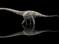
Dinosaur DNA and Proteins Found in Fossils, Paleontologists Claim

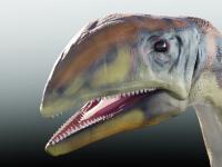
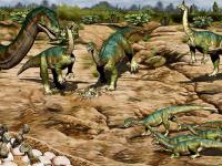
In the 1980s, paleontologists found a dinosaur nesting ground with dozens of nestlings in northern Montana and identified them as Hypacrosaurus stebingeri, a species of herbivorous duck-billed dinosaur that lived some 75 million years ago (Cretaceous period). Now, a team of researchers from the United States, Canada, and China has investigated molecular preservation of calcified cartilage in one of the Hypacrosaurus stebingeri nestlings at the extracellular, cellular and intracellular levels. They’ve found chemical markers of DNA, preserved fragments of proteins and chromosomes in the dinosaur chondrocytes (cartilage cells). The findings further support the idea that these original molecules can persist for tens of millions of years.
“The skull bones of baby dinosaurs are not fused when they hatch, but instead, some of them have cartilaginous plates that fuse later as bone forms in the spaces between them,” said Dr. Alida Bailleul, a paleontologist in the Institute of Vertebrate Paleontology and Paleoanthropology and the Center for Excellence in Life and Paleoenvironment, Chinese Academy of Sciences.
“Seeing exquisitely preserved microscopic structures that resembled the specific cell types found only in cartilage, and which would have been present in the living organism in these tissues, led us to hypothesize that cellular preservation may have extended to the molecular level.”
Dr. Bailleul and colleagues performed immunological and histochemical analyses of tissues from the skull of the Hypacrosaurus stebingeri hatchling and compared the results to those from an emu skull at a similar stage of development.
“Bird skulls ossify, or harden, in the same pattern as this hadrosaur’s skull would have, and primitive birds (ratites) like emus are the closest relatives we have alive today to non-avian dinosaurs,” said Professor Mary Schweitzer, a researcher at North Carolina State University, North Carolina Museum of Natural Sciences and the University of Lund.
The cartilaginous tissues and chondrocytes from the dinosaur skull reacted with antibodies to collagen II, but the surrounding bone did not react with collagen II antibodies.
This is significant because collagen II is found only in cartilage, while collagen I dominates in bone.
Comparing the results to the emu confirmed the findings.

“These tests show how specific the antibodies are to each type of protein, and support the presence of collagen II in these tissues,” Professor Schweitzer said.
“Additionally, bacteria cannot produce collagen, which rules out contamination as the source of the molecules.”
The scientists also tested the microstructures for the presence of chemical markers consistent with DNA using two complementary histochemical stains that bind to DNA fragments within cells: 4′,6′-diamidino-2-phenylindole dihydrochloride and propidium iodide.
These chemical markers reacted with isolated cartilaginous cells, supporting the idea that some fragmentary DNA may remain within the cells.
“We used two different kinds of intercalating stains, one of which will only attach to DNA fragments in dead cells, and the other which binds to any DNA,” Professor Schweitzer said.
“The stains show point reactivity, meaning they are binding to specific molecules within the microstructure and not smeared across the entire ‘cell’ as would be expected if they arose from bacterial contamination.”
“Although bone cells have previously been isolated from dinosaur bone, this is the first time that cartilage-producing cells have been isolated from a fossil,” Dr. Bailleul said.
“It’s an exciting find that adds to the growing body of evidence that these tissues, cells and nuclear material can persist for millions — even tens of millions — of years.”
The findings were published in the journal National Science Review.
_____
Alida M. Bailleul et al. Evidence of proteins, chromosomes and chemical markers of DNA in exceptionally preserved dinosaur cartilage. National Science Review, published online January 12, 2020; doi: 10.1093/nsr/nwz206
Source: www.sci-news.com/